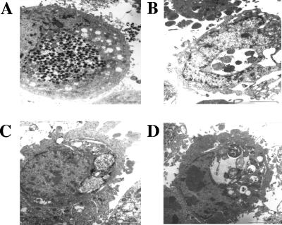
FIG. 3.
Transmission electron microscopy of chlamydial inclusions in acute and persistent infection at 48 h p.i. (A) HEp-2 monolayers infected with C. psittaci DC15 without persistence inducer. A large inclusion, with numerous EBs, numerous intermediate condensing bodies, and few RBs is shown. (B) Persistent infection at iron depletion (150 μM DAM). A small inclusion containing only a few RBs is shown. (C) Persistent infection after treatment with 200 U/ml penicillin G. An inclusion densely packed with RBs of variable size and shape is shown. Two RBs are severely enlarged. Note the electron-dense depositions at the outer membranes of RBs. (D) Persistent infection upon exposure to IFN-γ (240 U/ml). A small inclusion with large RBs at the periphery and amorphous material in the center is shown. RBs are highly variable in size, shape, and appearance. Bars, 2 μm.

