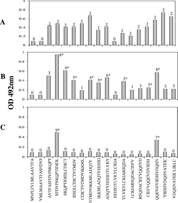
FIG. 2.
Mapping analysis using peptides spanning the entire sequence of the protein the FhSAP-2. For this experiment ELISA plates were coated with 20 μg/ml per well. Rabbit and mouse sera were tested at dilutions of 1:200. To visualize specific peptide-antibody reactions, peroxidase-labeled anti-species immunoglobulin G (Bio-Rad Laboratories, Hercules, CA) conjugate, diluted 1:5,000, was used. Individual peptides were tested with two specific anti-FhSAP-2 sera (A), eight sera from rabbits at 12 weeks postinfection with Fasciola (B), and nine sera from mice at 9 weeks postinfection with S. mansoni (C). Serum was considered positive when its OD at 492 nm exceeded the previously established cutoff value (cutoff, 0.19), which was calculated as the mean absorbance plus two standard deviations of the negative rabbit or mouse sera. Numbers above columns indicate the number of sera reacting with a given peptide. An asterisk over the bar indicates statistical difference (P > 0.05) between mean absorbance values with infected sera and negative sera for each specific peptide.

