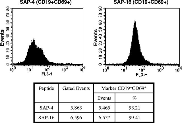
FIG. 4.
Peptides SAP-4 and SAP-16 contain, respectively, the immunodominant B-cell epitopes 21SKQPTIDIDL30 and 76QQEVDYIIDH85. They were conjugated with keyhole limpet hemocyanin, emulsified in Freund's adjuvant, and injected separately into two mice. Splenocytes were isolated from each immunized animal and cultured in triplicate at 106 cells/well in 200 μl of RPMI-1640 medium supplemented with 10% fetal bovine serum and 1% penicillin-streptomycin and stimulated with 5 μg/ml of each peptide. Cells were incubated for 48 h at 37°C under 5% CO2 atmosphere. After incubation, cells were centrifuged, washed with PBS, stained with anti-CD19-FITC or anti-CD69-PE-Cy7 (BD Pharmigen, San Diego, CA) monoclonal antibodies, and analyzed by fluorescence-activated cell sorting using a FACSort (Becton and Dickinson, San Jose, CA) instrument. Splenocytes were identified by their characteristic appearance on a dot plot of forward scatter versus side scatter. Splenocytes stained with FITC- or PE-Cy7-labeled antibodies were detected by FL1 or FL3, respectively. B-cells were gated on the basis of CD19 staining. The results were reported as the percentage of positive cells within this gate that appear activated.

