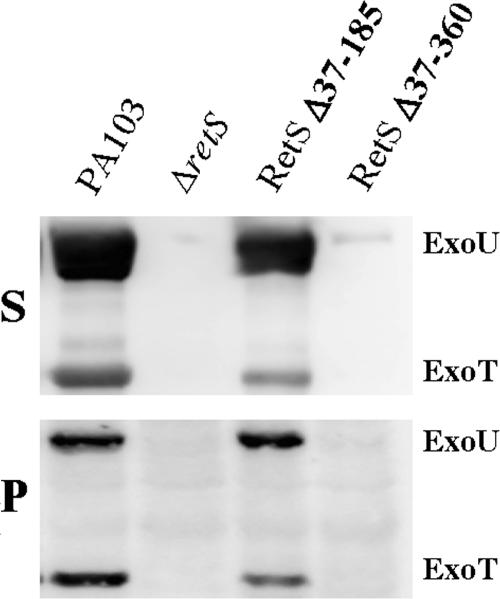
FIG. 5.
In vitro T3S of ExoU and ExoT does not require the periplasmic domain of RetS. Bacteria were grown overnight in T3S-inducing medium at 30°C. Samples were normalized to bacterial counts (5 × 10⁸/lane) or total protein (15 μg/lane) for supernatant (S) and lysate (P) samples, respectively. ExoU and ExoT were detected by Western blotting using a polyclonal antibody to the T3S effectors as described in Materials and Methods. The positions of ExoU and ExoT are indicated. The wild-type copy of retS and truncations of retS are expressed as single-copy chromosomal integrates under the control of the native retS promoter in a ΔretS strain.

