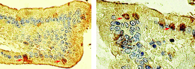
FIG. 2.
Immunohistochemistry performed on small intestinal sections of a SCID mouse (left) and a nude rat (right) using E. bieneusi-specific antibody, showing the presence of E. bieneusi forms within infected cells. The animals were inoculated with 106 spores each and were euthanized week 8 (left) and week 12 (right) after challenge (ABC method, DAB chromogen, hematoxylin counterstain; magnification, ×400). Arrows indicate infected cells.

