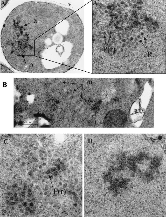
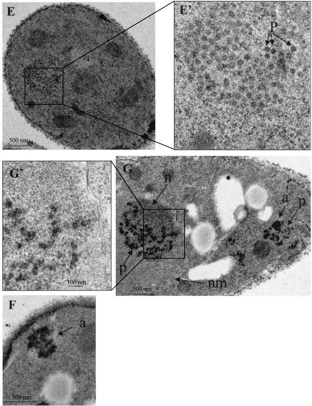
FIG. 5.
Electron micrographs of S. pombe cells expressing Tf1-WT (A, A', B, C, and D), Tf1-PRfs (G and G'), Tf1-ΔA (F), and Tf1-ΔD (E and E'). A', E', and G' are higher magnifications of the images shown in A, E, and G, respectively. a, aggregates of Tf1 material; P(r), particles with dense rings; P, particles without rings; m, membranes; n, heterochromatin; nm, nuclear membrane.


