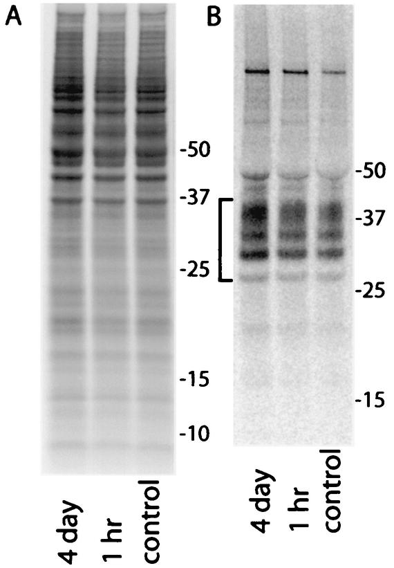
FIG. 3.
Phosphor autoradiographic images of 35S metabolic labeling of total proteins (A) and PrP-sen (B) in ScNB cells after pretreatments with 1 μM curcumin. Duplicate cultures were seeded and grown to confluence as was done in experiments such as those presented in Fig. 2. Curcumin was added to the culture medium either 4 days or 1 h prior to labeling of the cells at confluence with [35S]methionine. Curcumin was also maintained at the same concentration in the labeling medium. (A) Five-microliter aliquots of 1-ml cell lysates were run directly on the SDS-PAGE gel, and the remainder of each lysate was used for the immunoprecipitation of the 35S-PrP-sen samples shown in panel B. The bracket on the left in panel B marks PrP bands. Except for the test compound, the experimental protocols were the same as those described previously (5).

