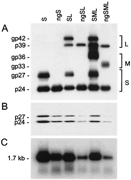
FIG. 2.
Production of N-linked-glycan-defective HDV particles. Culture fluids from HuH-7 cells were harvested on days 6, 9, and 12 after transfection of 106 cells with a mixture of 1 μg of HDV recombinant pSVLD3 plasmid DNA and 2 μg of pT7HB2.7 or mutant plasmid DNA coding for wt or unglycosylated HBV envelope proteins, respectively. Particles from the culture fluids were concentrated and assayed for the presence of HBV envelope proteins (A) or delta proteins (B) after electrophoresis on a 12% acrylamide gel, transfer to a polyvinylidene difluoride membrane, and immunodetection with a rabbit anti-HBsAg antibody (1:1,000 dilution) or rabbit anti-HDAg antibody (1:200 dilution). Rabbit immunoglobulin G was then probed with horseradish-labeled mouse anti-rabbit antibodies and a chemiluminescent substrate. Light emission was detected by exposure to Biomax ML film (Kodak). The molecular masses of the S, M, and L HBV envelope proteins and delta proteins are indicated at the left side of each panel. The size markers were prestained proteins (Amersham), measured in kilodaltons (e.g., gp42, glycoprotein with a molecular size of 42 kDa). Sedimented particles from the culture medium were also assayed for the presence of HDV RNA after RNA extraction, gel electrophoresis, and Northern blot hybridization by using a genomic strand-specific 32P-labeled HDV RNA probe (C). The size in kilobases of HDV genomic RNA is indicated.

