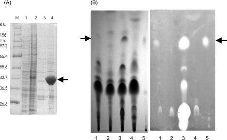
FIG. 3.
Overexpression of His6-tagged hemA-asuA in S. nodosus and its effect on asukamycin production. (A) The SDS-PAGE gel analysis (10% gel; 20 μg of the protein per lane was applied) shows cell extracts of S. nodosus carrying pIJ622 vector as a negative control (lane 1) or pALS4 plasmid (lane 2). The extracts from 1.5 ml of culture were subjected to affinity purification using TALON resin according to the manufacturer's instructions for native purification. The results are shown in lanes 3 (control) and 4 (His6-ALAS). The position of the ALAS band is indicated by an arrow. The molecular sizes of the marker (M) proteins are indicated. (B) The presence of the antibiotic in the cultivation broth was detected by TLC and bioassay against Bacillus subtilis 0453-52-9 (Difco) grown on a nutrient agar plate (CM0003; Oxoid). The left part shows a chromatogram visualized under UV light (254 nm); the right part represents the bioassay (chromatogram was laid on the surface of the agar plate for 15 min, and the plate was incubated overnight at 37°C). Into each lane, 10 μl of the extract was applied. The extracts were prepared from the following cultures: lane 1, parental S. nodosus strain; lane 2, MIP12 mutant; lane 3, MIP12 × pALS4; lane 4, MIP12 mutant supplemented with 20 μg/ml ALA. Asukamycin (10 μg) was applied as a control to lane 5. The position of asukamycin is indicated by an arrow. (The inhibition zone in the start of lane 3 is caused by thiostrepton, which was coextracted with asukamycin from the supplemented medium.)

