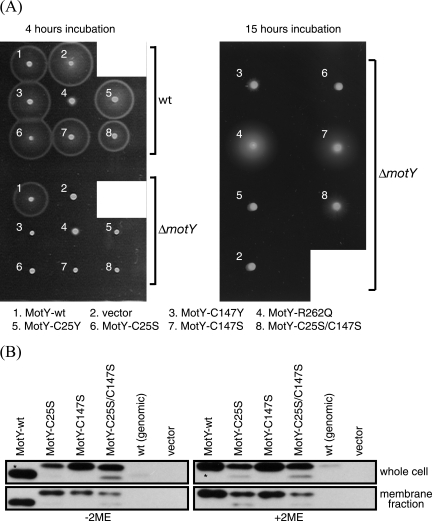
FIG. 1.
Characterization of MotY mutants. (A) VPG (1% polypeptone, 0.4% K2HPO4, 3% NaCl, and 0.5% [wt/vol] glycerol) plates containing 0.25% agar and 100 μg/ml kanamycin were inoculated with fresh colonies of the wild-type strain (VIO5) or the ΔmotY mutant strain (GRF2) expressing the indicated proteins from plasmids. They were incubated at 30°C for 4 h or 15 h. The plasmids and strains used are listed in Table 1. (B) Whole-cell lysates and the membrane fractions of GRF2 cells (ΔmotY) expressing the indicated proteins from plasmids and the wild-type strain [VIO5; wt (genomic)] were prepared and subjected to sodium dodecyl sulfate-polyacrylamide gel electrophoresis and immunoblotting as described previously (35) using anti-MotY antibodies (MotYB0079) in the absence (left) or the presence (right) of 2-mercaptoethanol (2ME). The asterisks (upper left and upper right) indicate that those samples were 1/10 the volume of the other samples. The anti-MotY antibody was raised against purified MotY produced from pKJ503 (25), which encoded C-terminally histidine-tagged MotY.

