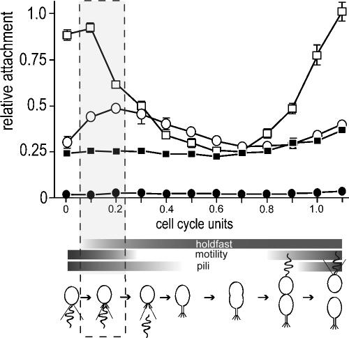
FIG. 1.
Surface attachment during the C. crescentus cell cycle. SW cells of the CB15 wild type (ATCC 19089) (open squares) and isogenic ΔpilA (open circles), ΔflgFG (closed squares), and ΔCC2277 (closed circles) mutants were purified and suspended in fresh peptone-yeast extract medium (per liter, 2.0 g of peptone and 1.0 g of yeast extract). Aliquots were removed from synchronized cultures throughout the cell cycle at 15-minute intervals, transferred to microtiter plates, and allowed to bind to the plastic surface for 15 min. Attachment was quantified by crystal violet staining according to the method of O'Toole and Kolter (19). The surface exposure and activity of polar organelles are indicated with horizontal bars below the time scale. Appearance and disappearance of pili were taken from Sommer and Newton (26). Motility was monitored microscopically throughout the cell cycle, and the presence of a polar holdfast was determined by fluorescent staining as described in the text. Cell cycle progression is indicated schematically below the graph. The time window of development during which flagellum, pili, and holdfast are exposed concomitantly at the same cell pole is boxed.

