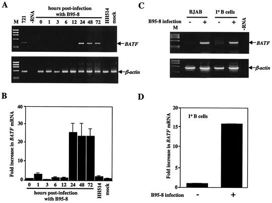
FIG. 2.
BATF mRNA is induced in human B cells following infection with EBV. (A) RT-PCR analysis of total RNA isolated at the indicated times (hours) from BJAB cells infected with B95-8 EBV or transformation-defective HH514 EBV. Amplification of BATF from the 721 B-cell line provided a positive control for expression. No RNA (−RNA) and RNA from mock-infected BJAB cells (mock) served as negative controls. ØX174 HaeIII DNA (M) served as a molecular weight marker. Each RT reaction was PCR amplified with primers for β-actin to control for sample integrity and amount. (B) Semiquantitative RT-PCR of the RT samples shown in panel A was performed as described in Material and Methods. The fold increase in BATF mRNA expression was quantified following the densitometry of ethidium bromide-stained gels. The experiment was performed three independent times with error bars indicating the standard error of the mean for each averaged set of values. (C) Primary (1° B) cells were isolated from human blood and mock infected (−) or infected with B95-8 EBV (+) as described in Materials and Methods. Total RNA from the primary B cells or control BJAB cells was isolated and examined for BATF mRNA expression by RT-PCR (upper panel). The amplification of β-actin served as the positive control. A no-RNA (−RNA) reaction served as the negative control. ØX174 HaeIII DNA (M) provided a marker for molecular weight. (D) Semiquantitative RT-PCR was performed on the 1° B-cell samples, and the fold increase in BATF mRNA was quantified as described for panel B.

