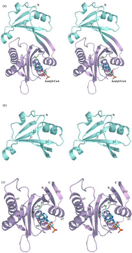
FIG.2.
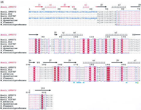
Three-dimensional structure of E. coli WecD. (a) Stereo view of the WecD monomer. The domains are differently colored; N-terminal domain is shown in cyan and the C-terminal GNAT domain in light pink. (b) The N-terminal domain with secondary structure elements labeled. (c) The C-terminal domain with secondary structure elements labeled. These and subsequent figures were prepared using PyMol (www.pymol.org). (d) Sequence alignment of selected WecD sequences, highlighting the N-terminal extended sequence present in WecD from E. coli CFT073 and Salmonella enterica (blue). The potential general acid, Tyr 208, is highlighted as a green star, and other AcCoA-binding residues are highlighted by cyan squares. Sequence alignment was carried out using ClustalW (7) and formatted using ESPript (23).


