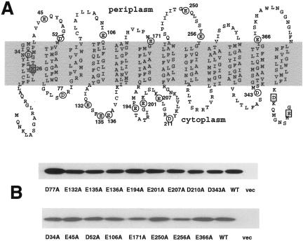
FIG. 1.
Neutralization of individual acidic residues in MdfA. (A) Acidic residues are marked on the secondary structure model of MdfA. Circled residues were analyzed in this study. Squares indicate residues that were analyzed previously (2, 4, 6). (B) Membranes were prepared from cells expressing the indicated His6-tagged mutants and analyzed by Western blotting. WT, wild type; vec, vector.

