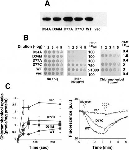
FIG. 3.
Characterization of MdfA mutants at positions D77 and D34. (A) Membranes were prepared from cells expressing the indicated His6-tagged mutants and analyzed by Western blotting. (B) E. coli was transformed with empty vector (vec) or plasmids encoding wild-type MdfA (WT) or the indicated MdfA mutants. Cells were diluted as marked [dilution (-log)] and spotted onto LB agar plates with or without the test drug, and plates were visualized after ∼20 h at 37°C. (C) Uptake of [3H]chloramphenicol by cells expressing the indicated mutants was assayed by rapid filtration (left panel). The experiment was repeated at least three times. The results shown are the average of triplicates. For the right panel, EtBr efflux was assayed as described in the legend of Fig. 2. a.u., arbitrary units.

