FIG. 1.
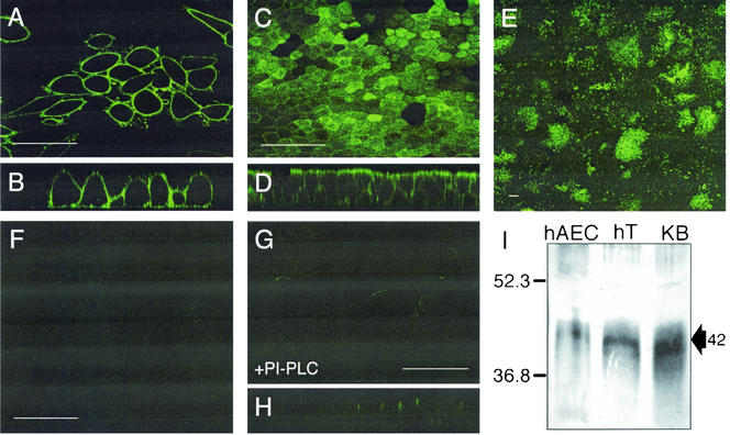
FRα expression in primary cultures of human airway epithelia. Cells were fixed and incubated with an FRα-specific monoclonal antibody, followed by addition of an anti-mouse FITC-conjugated secondary antibody. KB cells were viewed by using confocal microscopy from an en face (A) or vertical (B) section. Primary cultures of airway epithelia were also viewed en face at high power (C) and from a vertical section (D), as well as en face at low power (E). To confirm antibody specificity, an isotype control primary antibody was used (F). Primary cultures of human airway cells were also imaged following PI-PLC treatment to confirm enzyme function (en face [G] and vertical [H] sections). (I) Western blot of indicated protein samples was conducted by using the same FRα-specific monoclonal antibody followed by an anti-mouse alkaline phosphatase-conjugated secondary antibody. The expected 42-kDa band is indicated with an arrow. hAEC, human airway epithelial cell; hT, human trachea; KB, KB cell line. Scale bars = 50 μm (A, C, F, and G) or 100 μm (E).

