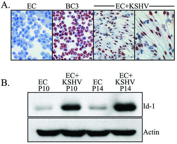
FIG. 2.
(A) Immunostaining of KSHV-infected ECs for LANA. Note the intense positive red reaction product in the nuclei of the KSHV-infected ECs (EC+KSHV). KSHV-infected ECs were cultured in LabTek chambers to preserve their spindle-shaped morphology during staining. Normal ECs (negative control) and BC-3 (positive control) cells are cytospin preparations and do not show cellular morphology. (B) Western blot analysis of Id-1 expression in ECs and KSHV-infected ECs (EC+KSHV). KSHV-infected cells expressed significantly higher levels of Id-1 than paired, control ECs. KSHV-infected ECs at passage 10 (P10) and P14 exhibited 11.3- and 25.3-fold increases, respectively, in Id-1 expression compared to Id-1 expression of ECs at the same passage number. The blot was probed for expression of Id-1 and reprobed for actin to demonstrate equal loading of the proteins. A representative experiment is shown. At least three independent experiments were performed and gave similar results.

