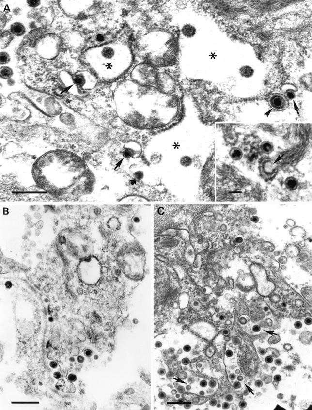
FIG. 6.
Secondary envelopment of nucleocapsids and exocytosis of virions. (A) Budding of intracytoplasmic nucleocapsids into cytoplasmic vesicles (arrows) resulted in mature virions inside exocytic vesicles (arrowhead). Note the presence of L particles within distended RER profiles (asterisks) and the formation of an intravesicular L particle (short arrow). The inset shows naked intracytoplasmic nucleocapsids adjacent to a RER profile marked with electron-dense granules (arrow) similar to those found after completion of the fusion event between the envelope of intracisternal L particles and the organelle membrane. (B and C) Simultaneous egress of virions and L particles at the basolateral membranes of epithelial cells. (C) Exocytosis of vesicles containing both virions and L particles (arrows). Bars, 500 nm and 100 nm (inset in panel A).

