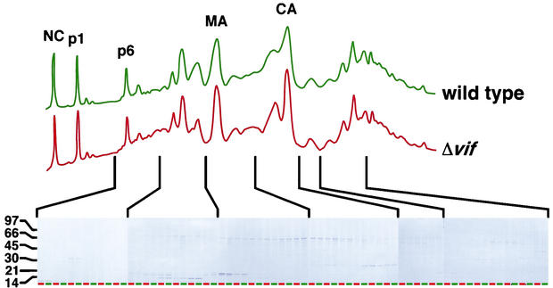
FIG. 5.
RP-HPLC examination of wild-type and Δvif HIV-1 virions. HIV-1YU-2 and HIV-1YU-2/Δvif produced in HUT8 cells were pelleted through sucrose cushions, disrupted, and separated by RP-HPLC. The identity of the peaks was determined by peptide sequencing and immunoblot analysis. The protein content of each fraction was visualized by SDS-PAGE and Coomassie blue staining. Adjacent lanes on the gels show matched fractions for wild-type (green) and Δvif (red) viruses.

