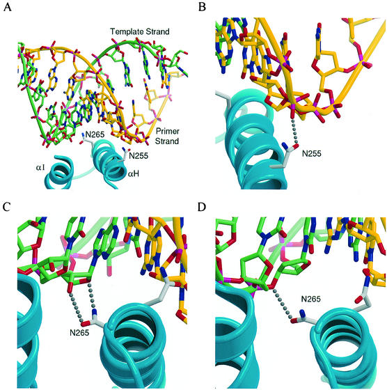
FIG. 3.
Interactions formed between the αH helix of the thumb subdomain with the minor groove of the template-primer. (A) Initial structure for the simulation of the N255D and N265D mutations with an RNA template. Helices H and I are shown as ribbons, with the side chains of N255 and N265 also indicated. The template and primer strands are also shown as ribbons in addition to a ball-and-stick representation. The backbone ribbon and carbons of the template strand are shown in light green, while those of the primer strand are shown in yellow. (B to D) Views of the RT-substrate interactions in the crystal structures of the wild-type RT with an RNA (40) and DNA (10) template, showing the N255 side chain hydrogen bonded to a phosphate on the DNA primer strand (B), the N265 side chain hydrogen bonded to O4′ and O2′ of a template ribose on the RNA template (C), and Asn-265 hydrogen bonded to O3′ of a template deoxyribose (D). Figures were generated by using MOLSCRIPT (29) and Raster3D (32).

