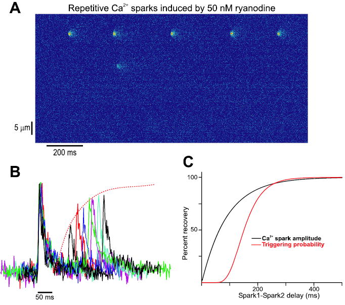
Figure 1.

Restitution of Ca2+ sparks in rat myocytes. (A) Addition of 50 nM ryanodine to a quiescent rat ventricular myocyte loaded with the Ca2+-sensitive dye fluo-3 causes a limited number of Ca2+ spark sites to display repetitive activity. Spark pairs derived from repeating sites such as the one near the top of the line scan image displayed can be analyzed to probe the time course of Ca2+ spark restitution. (B) Example of Ca2+ spark pairs recorded in rat ventricular myocytes after application of 50 nM ryanodine. Individual amplitudes of the initial Ca2+ sparks within each pair have been normalized so that spark pairs could be overlaid. Initial Ca2+ sparks display very similar time courses. The amplitude of the second spark in each pair, relative to the first, increases with an increase in the delay between sparks. (C) Ca2+ spark amplitude and triggering probability recovery functions derived from the analysis of spark pairs. Several potential regulatory factors, discussed in the text, could plausibly account for roughly 90 msec delay (measured at 50% recovery) between the two plots. The results shown have been replotted from the original data discussed in a recent study,23 with the permission of the authors.
