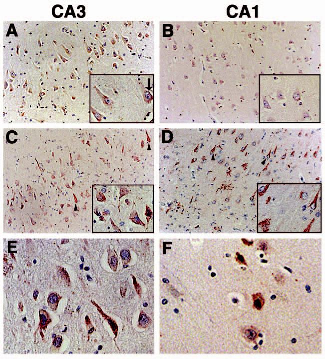
Figure 1.

p73 immunoreactivity in the CA3 and CA1 regions of the hippocampus using anti-p73 antibody H79. (A and B): p73 is located in neuronal cell bodies of the CA3 and CA1 hippocampal regions from control subjects. p73 is denoted in red and the sections counterstained with haematoxylin. Insets are high power magnification. (C and D): within the hippocampus of AD subjects, p73 is also detected in neurites and structures resembling neurofibrillary tangles (arrowheads). Each panel is ×200 magnification and the insets are ×400. (E and F): high power magnification (×400) of hippocampal pyramidal neurones immunolabelled for p73 in the CA3 (E) and CA1 (F) regions of AD brain. Panel (A) corresponds to case Control 8; panel (B) represents case Control 1; panel (C) is case AD 9; panel (D) is case AD 16; panel (E) is case AD 3; panel (F) is case AD 8 in Table 1. AD, Alzheimer disease.
