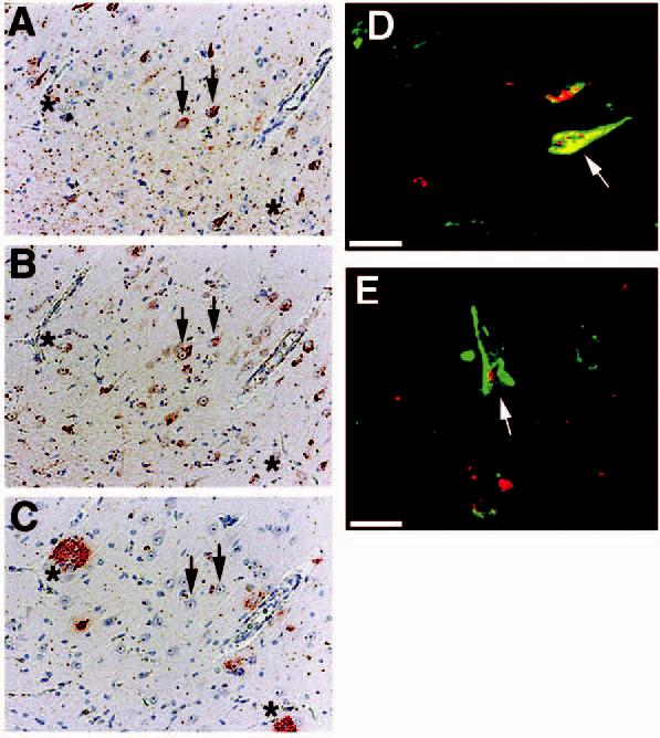
Figure 3.

p73 colocalizes to a subset of pyramidal neurones containing neurofibrillary tangles. (A–C): consecutive sections from Alzheimer disease (AD) hippocampus immunostained for Alz50 (A), p73 using the Ab-4 antibody (B), and beta-amyloid using 4G8 (C). Arrows indicate colocalization of p73 in tangle-bearing neurones and asterisks represent locations of amyloid-containing plaques. Magnification in panels A–C is ×200. (D and E): double-label confocal laser microscopy demonstrates colocalization of p73 (red) in tangle-bearing neurones (green) denoted using Alz50 antibody. Note that p73 as detected by the Ab-4 antibody is located in either the cytoplasm or nucleus (arrows) of these neurones. Bar corresponds to 20 μm in each panel. Panels (A–C) represent case AD 10, panel (D) represents case AD 13, and panel (E) represents case AD 6 in Table 1.
