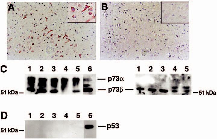
Figure 4.

Abundant p73 protein levels in the hippocampus. Consecutive sections from the CA1 region of the hippocampus were immunolabelled for p73 using the Ab-4 antibody (A) or p53 (B). While p73 immunoreactivity was evident, little p53 protein was observed in the hippocampus from control or Alzheimer disease (AD) subjects. Panels (A) and (B) represent case AD 8 in Table 1. Magnification for both panels is ×200. Insets are high power magnification of the same region of the section. (C) Immunoblot for p73 (left) and p73β (right) using tissue extracts from the hippocampus of control and AD subjects. Lanes 1 and 2 represent control subjects and lanes 3–5 represent AD subjects (AD cases 12, 13, and 7, respectively). Lane 6 is a nuclear extract from cultured neuroblastoma cells as a positive control for p73. H79 antibody was used to detect p73 and C20 was used to detect p73β. Equal protein was loaded into each lane. (D) p53 protein was below detectable limits within the same tissue extracts. Identical lane assignments as in panel C.
