
Figure 5. Hsp104 couples ATP hydrolysis to Sup35 prion fragmentation.
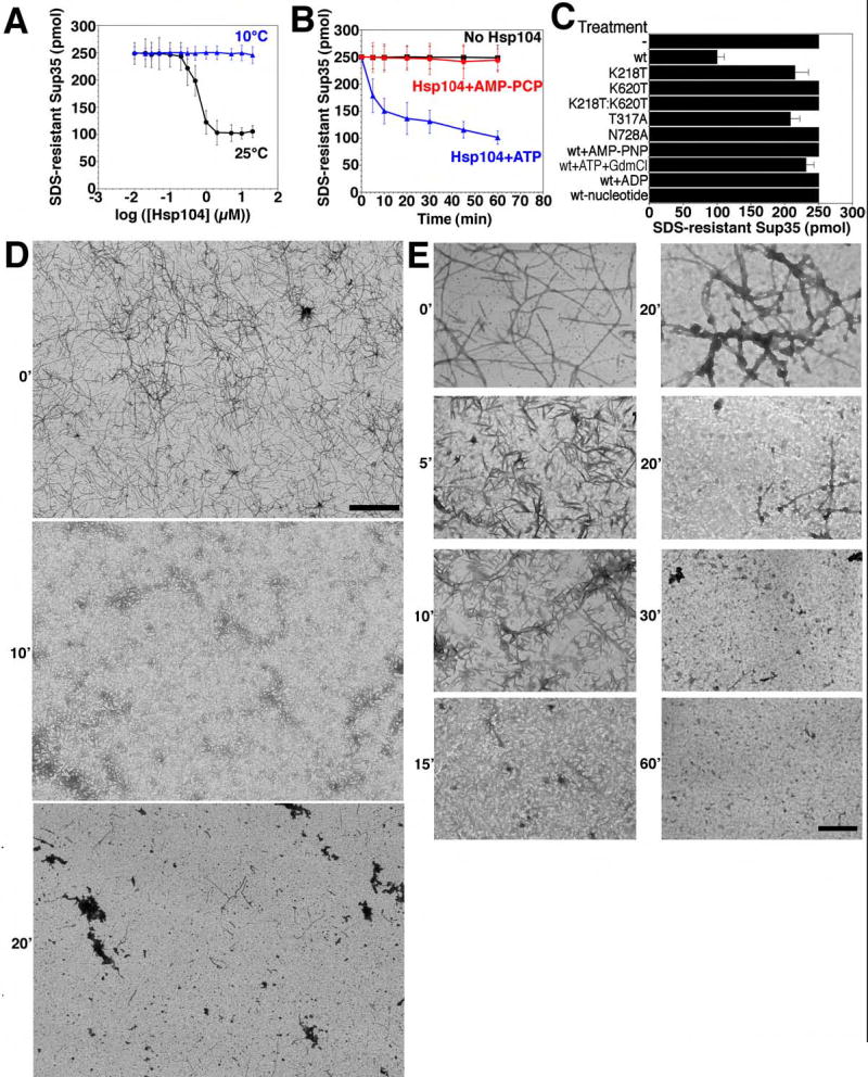
(A) Sup35 (2.5μM) was incubated for 8h with rotation (80rpm) to generate fibers and then incubated with Hsp104 (0.01–20μM) plus ATP (5mM) for 60min at 25°C (black) or 10°C (blue). Disassembly was assessed by the amount of SDS-resistant Sup35. Values represent means±SD (n=3).
(B) Kinetics of Hsp104 (2μM) induced Sup35 fiber (2.5μM monomer) disassembly in the presence of ATP (5mM) or the non-hydrolyzable analog AMP-PCP (5mM) (an alternative to AMP-PNP) at 25°C. Disassembly was monitored as in (A). Values represent means±SD (n=3).
(C) Sup35 fibers (2.5μM monomer) were treated at 25°C with the indicated Hsp104 proteins (2μM) plus ATP (5mM) or Hsp104 plus AMP-PNP (5mM), ATP (5mM) and GdmCl (20mM), ADP (5mM), or no nucleotide. Wt denotes wild type Hsp104. Disassembly was monitored by the amount of SDS-resistant Sup35. Values represent means±SD (n=3).
(D, E) Sup35 fibers (2.5μM monomer) were incubated with Hsp104 (2μM) plus ATP (5mM) for 0–60min at 25°C, and at various times reactions were processed for EM. Scale bars: 2μm (D), 0.25μm (E).