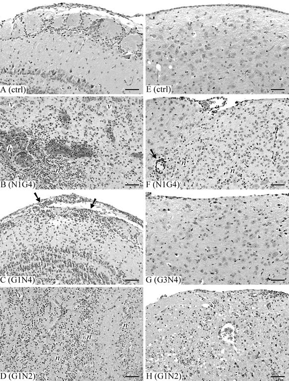
FIG. 3.
Lesion development in the olfactory bulb and brain tissue of mice at 7 days postinoculation with the rearranged viruses. Groups of four to seven mice were inoculated intranasally with 5,000 PFU of N1G4(wt), G1N2, G3N4, or G1N4 per animal. Control animals were given medium only. Brains were fixed in situ, sectioned, and stained with H&E. (A) Olfactory bulb of uninfected control. (B) Olfactory bulb of N1G4(wt)-infected animal showing necrotizing encephalitis with necrosis (n), hemorrhage (h), and neutrophil accumulation. v, viable tissue. (C) Olfactory bulb of mouse inoculated with attenuated G1N4 virus showing lymphocytic meningoencephalitis, with lymphocytic infiltration of leptomeninges and superficial neutrophils (arrows). (D) Olfactory bulb of mouse given virulent G1N2 virus showing severe extensive necrotizing encephalitis (n). (E) Medulla of a sham-inoculated control. (F) Medulla of mouse given N1G4(wt) virus showing mild necrotizing encephalitis, with lymphocytic perivascular cuffing (arrow) and inflammatory cell infiltration and neuronal necrosis (n). (G) Medulla of mouse inoculated with attenuated G3N4 virus showing no abnormalities. (H) Medulla of mouse given virulent G1N2 virus showing necrotizing encephalitis with diffuse infiltration of neutrophils and lymphocytes. Bars, 65 μm.

