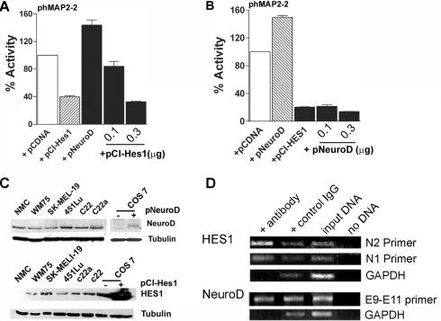
Figure 7.
HES1 is a dominant repressor of MAP2 promoter activity. (A) Melanoma cells were transfected with phMAP2-2 promoter-luciferase plasmid alone, or with Hes1 expression plasmid pCI-Hes1 or NeuroD expression plasmid pcDNA3-NeuroD-FLAG. In addition, 0.1 and 0.3 μg of Hes1 expression plasmid pCI-Hes1 was included in cells co-transfected with NeuroD. (B) c22a melanoma cells were transfected with phMAP2-2 promoter-luciferase plasmid alone, or with Hes1 expression plasmid pCI-Hes1 or NeuroD expression plasmid pcDNA3-NeuroD-FLAG. Cells in additional wells transfected with pCI-Hes1 also received 0.1 or 0.3 μg of pNeuroD plasmid. Equal amount of total DNA was used in all transfection. Luciferase activities were measured and the promoter activity in cells co-transfected with either pcDNA3-NeuroD or pCI-Hes1 alone or with increasing amounts of pCI-Hes1 or pcDNA3-NeuroD1, respectively, is shown as percentage (means ± SEM) of activity in cells transfected with phMAP2-2 and pcDNA. (C) Expression of NeuroD and HES1 in melanocytes and melanoma cells. Western blot analysis of NeuroD expression in control and pcDNA3-Neuro-FLAG plasmid transfected COS cells, neonatal foreskin melanocytes (NMC), primary melanoma cell line WM75 and various metastatic melanoma cell lines. Seventy-five micrograms of detergent soluble total cellular proteins were separated by SDS–PAGE and immunoblotted with polyclonal goat anti-mouse NeuroD (upper panel) and anti-mouse Hes1 antibody (lower panel), followed by HRP-conjugated secondary antibody. Protein bands were detected by chemiluminescence. Western blotting for α-tubulin is shown as a control for protein loading. (D) Chromatin immunoprecipitation assay. Sheared chromatin from melanoma cells was immunoprecipitated (IP) with appropriate antibody (anti-HES1, anti-NeuroD or control IgG) and antibody bound DNA was isolated according to the manufacturer's protocol (Active Motif, Carlsbad, CA). Immunoprecipitated DNA was used as template in PCR using N1 and N2 primer (for HES1), E9–E11 primer (for NeuroD). GAPDH primers were used as control.

