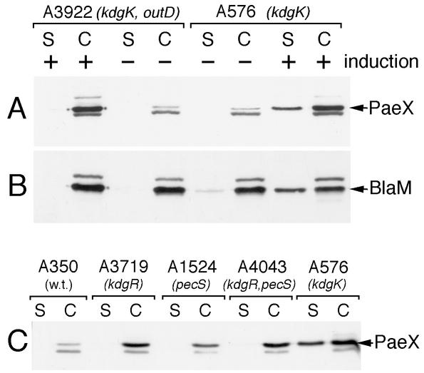
FIG. 6.
Cellular localization of PaeX. (A and B) E. chrysanthemi A576 (kdgK) and A3922 (kdgK outD) carrying the pT7-6 plasmid were grown in LB medium (−) or in LB medium supplemented with 0.2% galacturonate (+) until the early stationary phase. The culture supernatant (S) and whole-cell (C) fractions were separated by SDS-PAGE and analyzed by immunoblotting with either PaeX (A) or BlaM (B) antibodies. (C) The parental E. chrysanthemi strain A350 and different regulatory mutants were grown in LB medium supplemented with 0.2% galacturonate. The culture supernatant (S) and whole-cell (C) fractions were analyzed by immunoblotting. Positions of PaeX and BlaM are indicated by arrows.

