Figure 4.
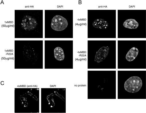
The 4xMBD protein is a sensitive stain for methyl-CpG-rich regions in fixed cells. Permeabilized and fixed mouse fibroblasts were incubated with 50 µg/ml 1xMBD (A) or 4 µg/ml 4xMBD (B), or their R22A mutant forms, and visualized with anti-HA plus Alexa-594-conjugated anti-mouse antibodies. Specific staining is seen at heterochromatic DAPI bright spots with the wild-type probe but not with their mutant counterparts. Weak staining by the 4xMBD–R22A mutant protein is sometimes detected, but does not co-localize with DAPI bright spots. Staining shows improved resolution with 4xMBD compared to 1xMBD and requires >10-fold less protein. (C) Metaphase spreads from mouse fibroblasts were incubated with 4 µg/ml 4xMBD and visualized with anti-HA plus Alexa-594-conjugated anti-mouse antibodies. DNA is counterstained with DAPI. Intense staining of pericentromeric satellite DNA is apparent plus weaker staining of the chromosome arms.

