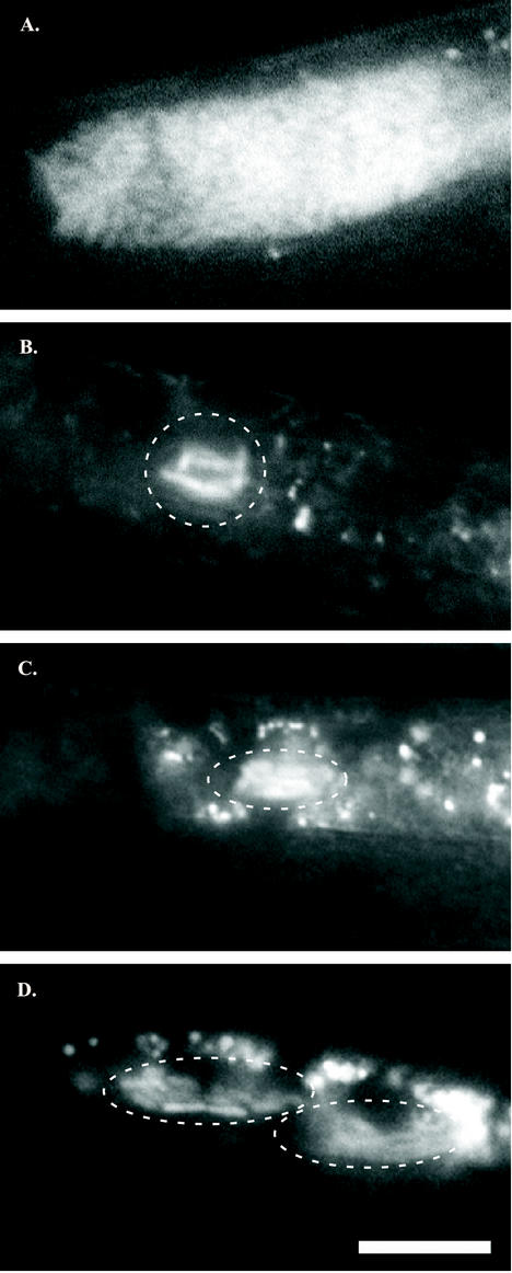
FIG. 1.
Bacterial colonization of immature IJs. Vesicles of mature IJs may contain 40 to >100 X. nematophila cells (A), whereas the vesicles of immature IJs contain only a few X. nematophila cells (indicated by enclosure in a dashed white line) (B to D). In panel B, six to seven rod-shaped X. nematophila cells cluster in close proximity to each other in the vesicle. The vesicle shown in panel C contains slightly more X. nematophila cells, and that in panel D contains even more than the IJ shown in panel B, but each has noticeably fewer than the full complement of cells found in a mature IJ (A). GFP-labeled X. nematophila cells were distinguished from nematode intestinal autofluorescence by virtue of bacterial cell shape and differential spectral emission under appropriate fluorescence filters (see Materials and Methods). In all images, nematodes are oriented with heads off the left side of the panel. Magnification, ×600. Bar, 10 μm.

