
Figure 4.
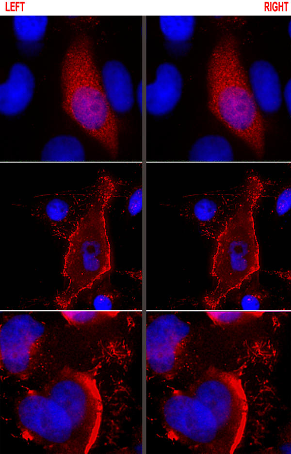
Ebola VP40 changes distribution during the course of infection. The stereoprojected images of MCF7 cells show that at the early phase of infection (top panel) VP40 is localized to homogeneously dispersed granules in the cytoplasm as well as in the nucleus. Later, at the initial phase of rounding up (middle panel), VP40 is almost exclusively membrane associated and it is present in extracellular filamentous structures as well. VP40 is also present in the virions that accumulate in large quantities around cells with pronounced cytopathic effects (bottom panel). DNA staining is blue.