
Figure 5.
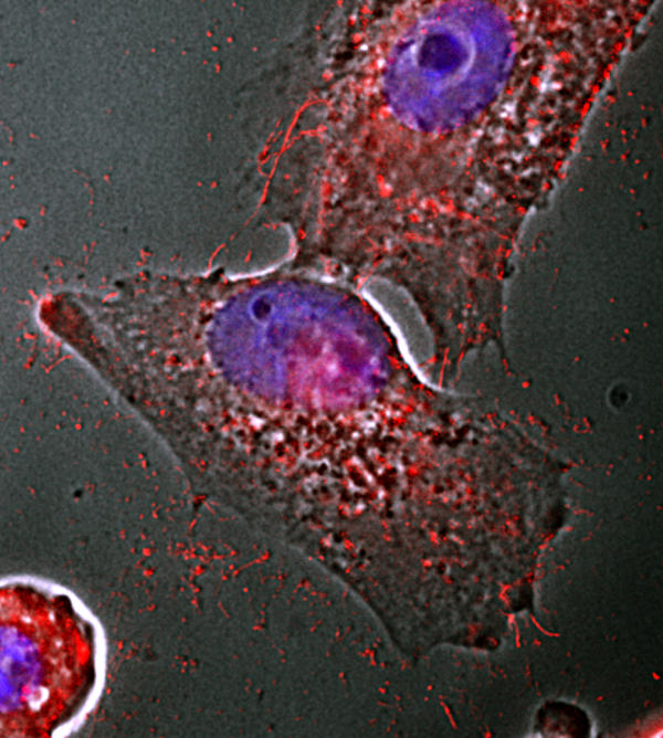
Release of filamentous Ebola virus particles from Vero E6 cells. VP40 staining (red) superimposed on a phasecontrast image of infected cells shows the appearance of extracellular virions even before the development of cytopathic effects. DNA staining is blue.