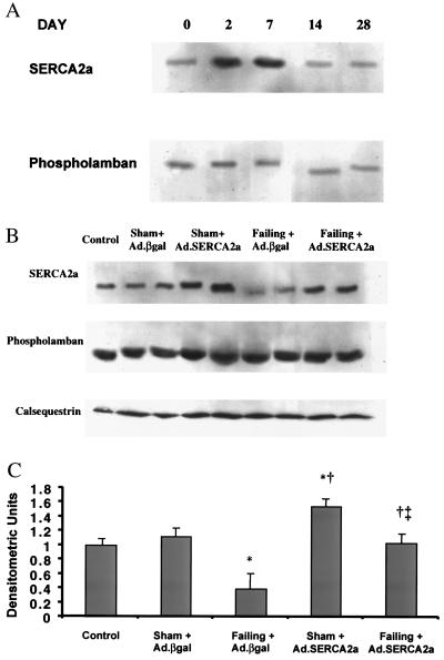
Figure 2.
(A) Immunoblots of SERCA2a and phospholamban at days 0, 2, 7, 14, and 28 after infection with Ad.SERCA2a in control rats. (B) Immunoblots of SERCA2a, phospholamban, and calsequestrin from crude membranes of left ventricles from control, sham rats infected with Ad.βgal, sham rat hearts infected with Ad.SERCA2a, failing rat hearts infected with Ad.βgal, and failing hearts infected with Ad.SERCA2a at day 2–3. (C) Protein levels of SERCA2a in preparations from sham rats infected with Ad.βgal (n = 7), sham rat hearts infected with Ad.SERCA2a (n = 8), preparations from failing rat hearts infected with Ad.βgal (n = 8), and preparations of failing hearts infected with Ad.SERCA2a at day 2–3 (n = 9). *, P < 0.05 compared with Sham + Ad.βgal; †, P < 0.05 compared with Failing + Ad.βgal; ‡, P < 0.05 compared with Sham + Ad.SERCA2a.

