
Figure 3.
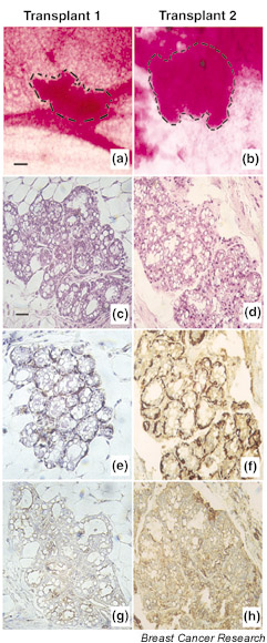
Histology and immunocytochemistry of two representative side population (SP) outgrowths. (a), (b) Wholemount morphology of carmine-stained outgrowths from transplanted mouse mammary side population cells. Lobuloalveolar structures indicated by dashed outlines. Bar = 750 μm. (c), (d) H & E-stained section of SP outgrowth. (e), (f) Immunocytochemical staining of the section of SP outgrowth for the myoepithelial cell marker α-smooth muscle actin. (g), (h) Immunohistochemical staining of the section of SP outgrowth for the luminal epithelial cell marker cytokeratin 19. (c)–(h) Bar = 350 μm.