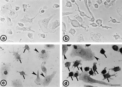
Figure 2.
Light microscopic immunocytochemical analysis of ABCG1 expression in cultured human macrophages. (a) Immunostaining of unloaded macrophages cultured for 4 days without addition of the secondary antibody (control 1). The absence of staining verifies the complete inhibition of endogenous peroxidases. (b) Macrophages incubated with acLDL for 2 days, stained without primary antiserum (control 2), indicating the complete blocking of nonspecific binding sites of the secondary antibody. (c) AcLDL-laden macrophages as in b, incubation with preimmune serum (control 3). Flattened cells (arrowheads) show less nonspecific labeling than smaller cells that extend discrete cellular processes (arrows). (d) Intracellular ABCG1 immunostaining in acLDL-laden macrophages. Note the intense staining in small macrophages (arrows) compared with large spread-out cells (arrowheads). A faint labeling in the perinuclear region (asterisks) is visible in flattened cells. Immunosignals were visualized by using the immunoperoxidase technique, and cell nuclei were counterstained with hematoxylin. (Bar = 20 μm.)

