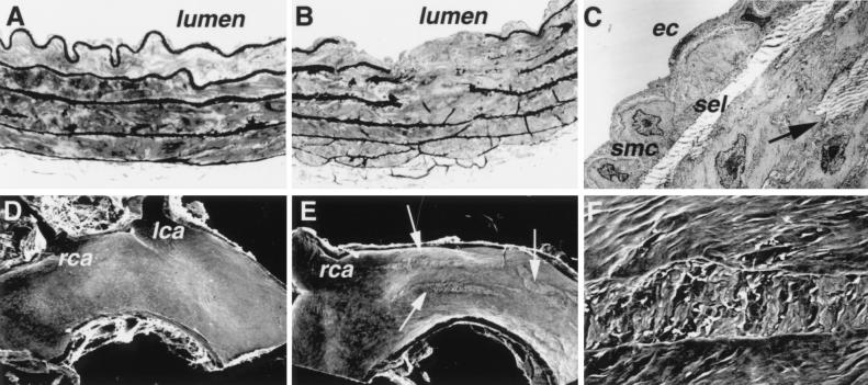
Figure 5.
Abnormalities in the aorta of Gulo −/− mice. (A) A cross section of the descending thoracic aorta from a wild-type mouse showing uninterrupted elastic laminae. Toluidine blue staining, light microscopy, ×400. (B) A cross section of the descending thoracic aorta from a Gulo −/− mouse with disrupted elastic laminae. Light microscopy, ×400. (C) TEM of the area adjacent to that shown in B. A single layer of mildly activated smooth muscle cells (smc) is present in the intima between endothelial cells (ec) and superficial elastic lamina (sel). A break in the elastica is marked by an arrow. Magnification: ×4,000. (D) Scanning electron microscopy of the aortic arch of a wild-type mouse showing a normal luminal surface. lca, left carotid artery; rca, right carotid artery. Magnification: ×20. (E) Scanning electron microscopy of the aortic arch of an Gulo −/− mouse showing multiple longitudinal surface defects (arrows). Magnification: ×20. (F) Higher magnification (×200) of the defect.

