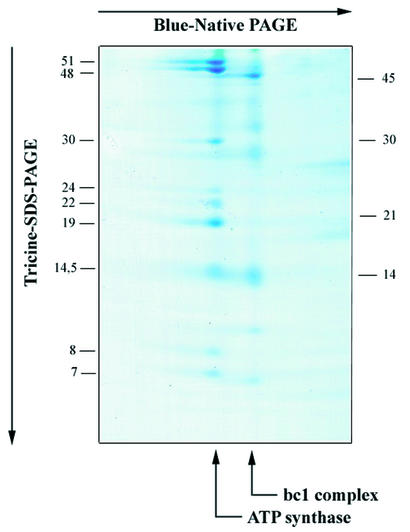
Figure 1.
Separation of ATP synthase subunits of S.ecuadoriensis. Membrane complexes were separated by blue-native polyacryl amide gel electrophoresis in the first dimension, and ATP synthase subunits were separated by Tricine–SDS polyacryl amide gel electrophoresis in the second dimension. Sizes of standard proteins are indicated in the right margin, and inferred sizes of ATP synthase subunits are given in the left margin. The protein complex from S.ecuadoriensis was identified by the characteristic subunit pattern, and sequence analysis of selected subunits using mass spectrometry (26).

