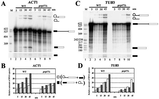
Figure 6.
In vitro splicing of ACT1 and TUB3 in wild-type and prp17::LEU2 extracts. (A) 32P-labeled ACT1 pre-mRNA was reacted with a wild-type extract at 23°C. Aliquots were withdrawn at the time points given above each lane (lanes 2–5). The RNA products were analyzed by denaturing PAGE. The RNA species lariat–exon 2, lariat intron, precursor, mRNA and exon 1 are indicated to the right. The splicing reaction performed with ACT1 substrate and extracts from prp17::LEU2 were run on lanes 6–9. (C) 32P-labeled TUB3 pre-mRNA substrate reacted with wild-type (lanes 2–5) or prp17::LEU2 extract (lanes 7–10). The reaction aliquots were withdrawn at the time points given above each lane. The RNA species were resolved on denaturing PAGE and are represented schematically on the right. (B and D) Quantitation of the splicing reactions 1 and 2 for the substrate ACT1 (B) or TUB3 (D). In each case, quantitations were done for reactions with wild-type extract and also prp17::LEU2 extracts. Open bars indicate the amounts of lariat–exon 2 plus exon 1 resulting from reaction 1 at the given time point. Filled-in bars indicate the amounts of lariat intron plus mRNA resulting from reactions 1 and 2 at the given time point. The quantitations of pre-mRNA, lariat–exon 2, lariat intron, mRNA and exon 1 were obtained through phosphorimager analysis of the gels. The levels of the RNA intermediates and product RNAs were normalized to the levels of precursor RNA in each lane.

