Abstract
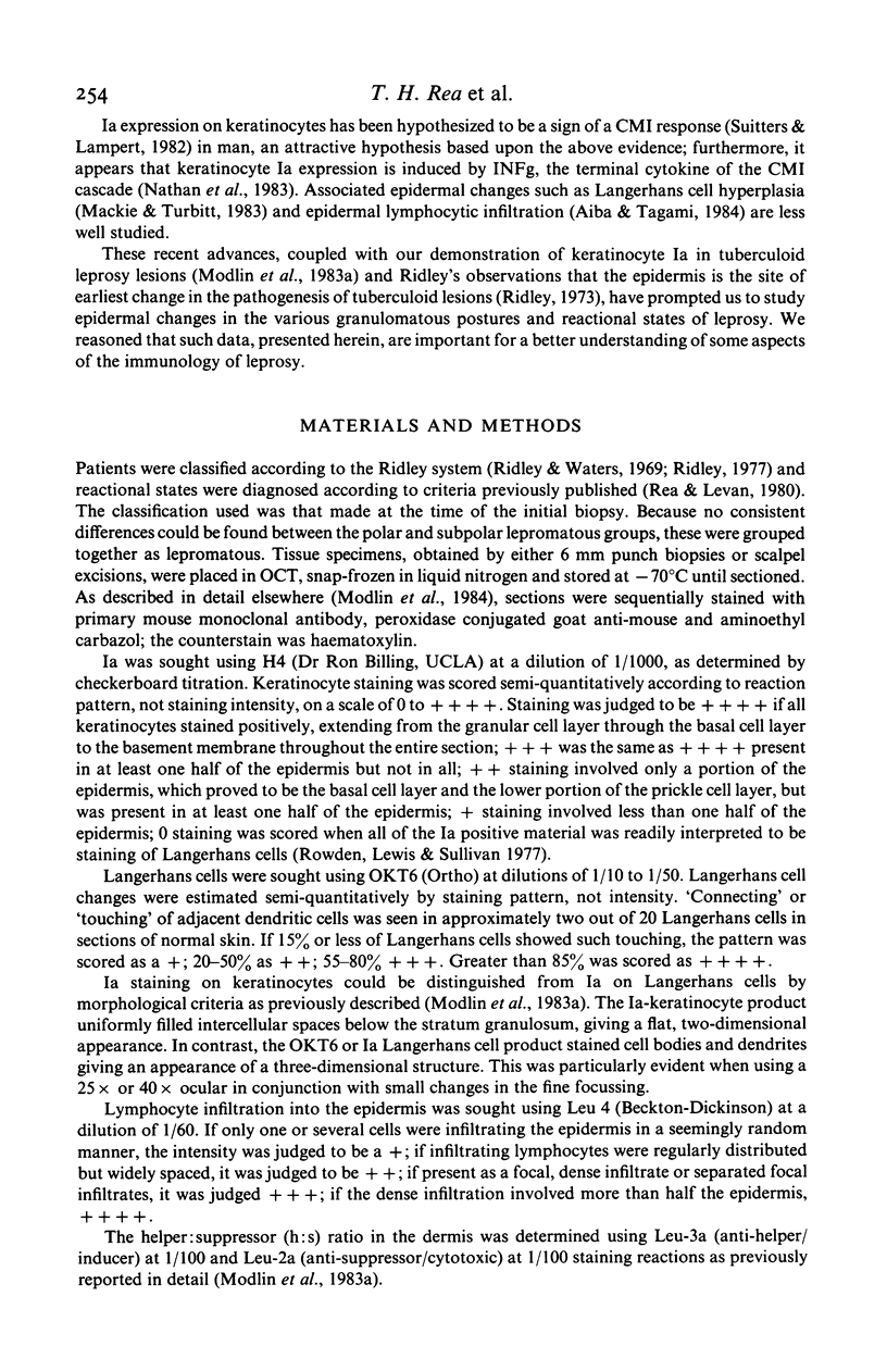
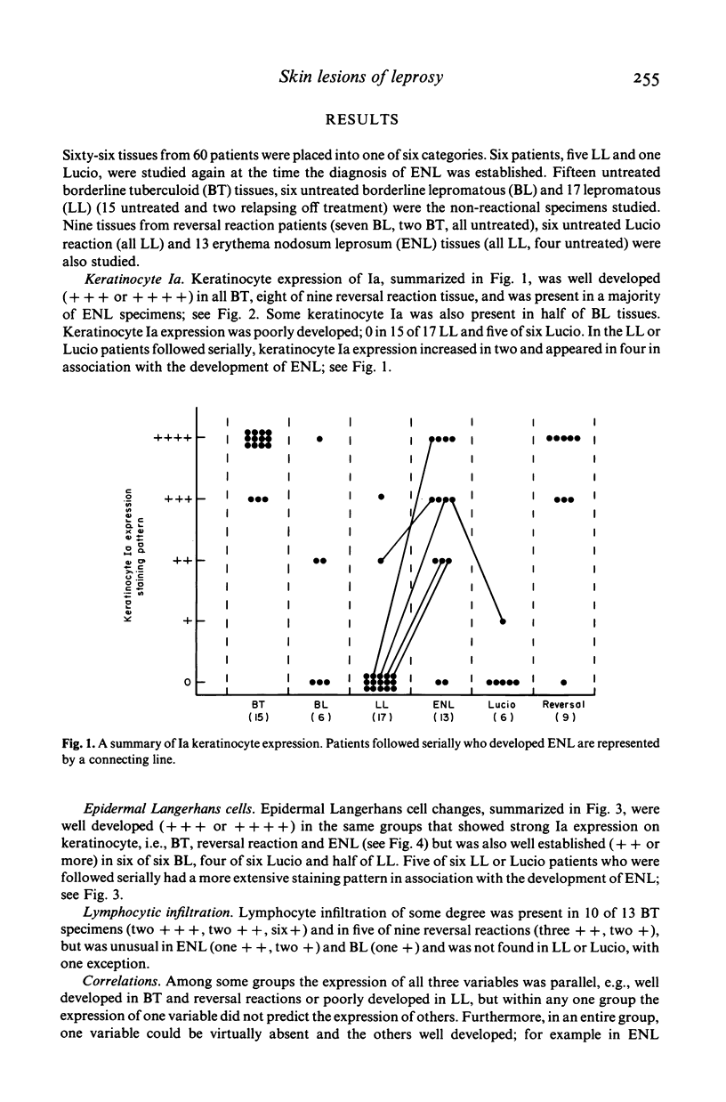
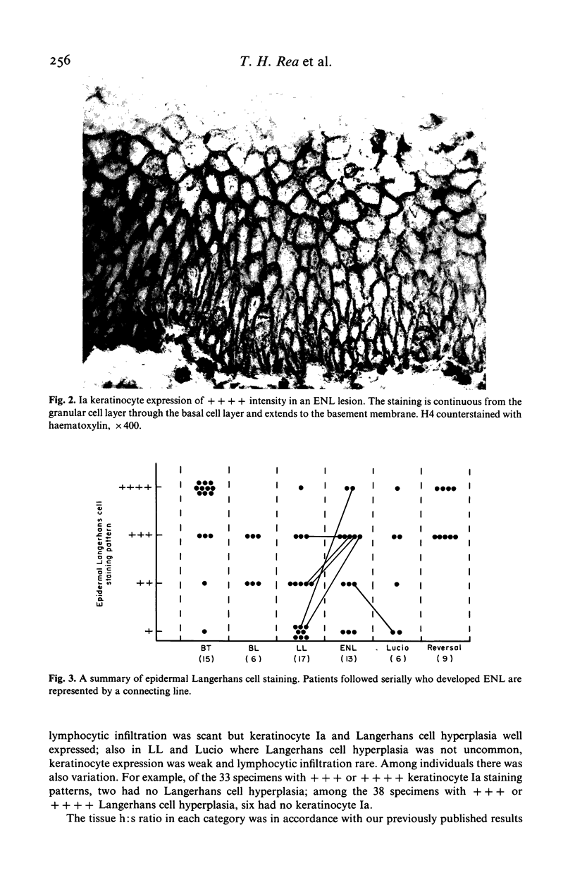
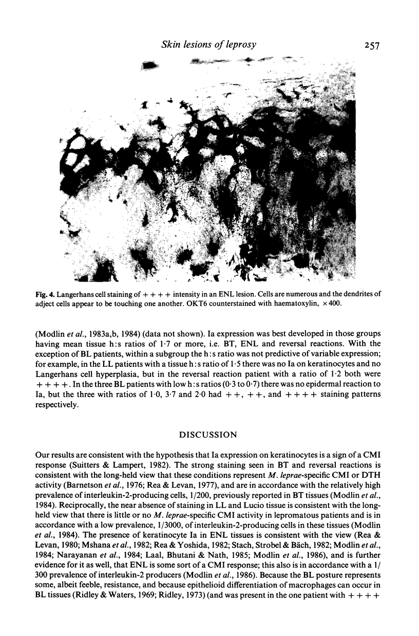
Epidermal changes, Ia expression on keratinocytes, Langerhans cell hyperplasia and lymphocyte infiltration were sought in skin lesions of leprosy: 15 borderline tuberculoid (BT), six borderline lepromatous (BL), 17 lepromatous (LL), 13 erythema nodosum leprosum (ENL), six Lucio reactions and nine reversal reactions. All three changes were well developed in BT and reversal reactions. ENL showed well developed keratinocyte Ia and Langerhans cell hyperplasia, but little lymphocytic infiltration. LL and Lucio tissues had some Langerhans cell hyperplasia but little or no keratinocyte Ia or lymphocytic infiltration. BL tissues were so diverse as to suggest two distinct subgroups. These findings are consistent with the hypothesis that keratinocyte Ia expression is an immunohistological sign of a cell-mediated immune (CMI) response. However, the Ia keratinocyte expression found in BL and ENL tissues appears contrary to the undifferentiated macrophages and numerous bacilli found in the lesions. Thus, if a sign of CMI, keratinocyte Ia expression is not a measure of the effectiveness of the response.
Full text
PDF
Images in this article
Selected References
These references are in PubMed. This may not be the complete list of references from this article.
- Aiba S., Tagami H. HLA-DR antigen expression on the keratinocyte surface in dermatoses characterized by lymphocytic exocytosis (e.g. pityriasis rosea). Br J Dermatol. 1984 Sep;111(3):285–294. doi: 10.1111/j.1365-2133.1984.tb04725.x. [DOI] [PubMed] [Google Scholar]
- Barnetson R. S., Bjune G., Pearson J. M., Kronvall G. Cell mediated and humoral immunity in "reversal reactions". Int J Lepr Other Mycobact Dis. 1976 Jan-Jun;44(1-2):267–274. [PubMed] [Google Scholar]
- Basham T. Y., Nickoloff B. J., Merigan T. C., Morhenn V. B. Recombinant gamma interferon induces HLA-DR expression on cultured human keratinocytes. J Invest Dermatol. 1984 Aug;83(2):88–90. doi: 10.1111/1523-1747.ep12262597. [DOI] [PubMed] [Google Scholar]
- Breathnach S. M., Katz S. I. Keratinocytes synthesize Ia antigen in acute cutaneous graft-vs-host disease. J Immunol. 1983 Dec;131(6):2741–2745. [PubMed] [Google Scholar]
- Katz S. I. The skin as an immunologic organ. A tribute to Marion B. Sulzberger. J Am Acad Dermatol. 1985 Sep;13(3):530–536. doi: 10.1016/s0190-9622(85)70195-8. [DOI] [PubMed] [Google Scholar]
- Laal S., Bhutani L. K., Nath I. Natural emergence of antigen-reactive T cells in lepromatous leprosy patients during erythema nodosum leprosum. Infect Immun. 1985 Dec;50(3):887–892. doi: 10.1128/iai.50.3.887-892.1985. [DOI] [PMC free article] [PubMed] [Google Scholar]
- Lampert I. A., Suitters A. J., Chisholm P. M. Expression of Ia antigen on epidermal keratinocytes in graft-versus-host disease. Nature. 1981 Sep 10;293(5828):149–150. doi: 10.1038/293149a0. [DOI] [PubMed] [Google Scholar]
- MacKie R. M., Turbitt M. L. Quantitation of dendritic cells in normal and abnormal human epidermis using monoclonal antibodies directed against Ia and HTA antigens. J Invest Dermatol. 1983 Sep;81(3):216–220. doi: 10.1111/1523-1747.ep12517998. [DOI] [PubMed] [Google Scholar]
- Modlin R. L., Gebhard J. F., Taylor C. R., Rea T. H. In situ characterization of T lymphocyte subsets in the reactional states of leprosy. Clin Exp Immunol. 1983 Jul;53(1):17–24. [PMC free article] [PubMed] [Google Scholar]
- Modlin R. L., Hofman F. M., Horwitz D. A., Husmann L. A., Gillis S., Taylor C. R., Rea T. H. In situ identification of cells in human leprosy granulomas with monoclonal antibodies to interleukin 2 and its receptor. J Immunol. 1984 Jun;132(6):3085–3090. [PubMed] [Google Scholar]
- Modlin R. L., Mehra V., Jordan R., Bloom B. R., Rea T. H. In situ and in vitro characterization of the cellular immune response in erythema nodosum leprosum. J Immunol. 1986 Feb 1;136(3):883–886. [PubMed] [Google Scholar]
- Mshana R. N., Haregewoin A., Harboe M., Belehu A. Thymus dependent lymphocytes in leprosy. I. T lymphocyte subpopulations defined by monoclonal antibodies. Int J Lepr Other Mycobact Dis. 1982 Sep;50(3):291–296. [PubMed] [Google Scholar]
- Narayanan R. B., Laal S., Sharma A. K., Bhutani L. K., Nath I. Differences in predominant T cell phenotypes and distribution pattern in reactional lesions of tuberculoid and lepromatous leprosy. Clin Exp Immunol. 1984 Mar;55(3):623–628. [PMC free article] [PubMed] [Google Scholar]
- Nathan C. F., Murray H. W., Wiebe M. E., Rubin B. Y. Identification of interferon-gamma as the lymphokine that activates human macrophage oxidative metabolism and antimicrobial activity. J Exp Med. 1983 Sep 1;158(3):670–689. doi: 10.1084/jem.158.3.670. [DOI] [PMC free article] [PubMed] [Google Scholar]
- Rea T. H., Levan N. E. Current concepts in the immunology of leprosy. Arch Dermatol. 1977 Mar;113(3):345–352. [PubMed] [Google Scholar]
- Rea T. H., Levan N. E. Variations in dinitrochlorobenzene responsivity in untreated leprosy: evidence of a beneficial role for anergy. Int J Lepr Other Mycobact Dis. 1980 Jun;48(2):120–125. [PubMed] [Google Scholar]
- Rea T. H., Yoshida T. Serum macrophage migration inhibition activity in patients with leprosy. J Invest Dermatol. 1982 Nov;79(5):336–339. doi: 10.1111/1523-1747.ep12500088. [DOI] [PubMed] [Google Scholar]
- Ridley D. S. The pathogenesis of the early skin lesion in leprosy. J Pathol. 1973 Nov;111(3):191–206. doi: 10.1002/path.1711110307. [DOI] [PubMed] [Google Scholar]
- Ridley D. S., Waters M. F. Significance of variations within the lepromatous group. Lepr Rev. 1969 Jul;40(3):143–152. doi: 10.5935/0305-7518.19690026. [DOI] [PubMed] [Google Scholar]
- Rowden G., Lewis M. G., Sullivan A. K. Ia antigen expression on human epidermal Langerhans cells. Nature. 1977 Jul 21;268(5617):247–248. doi: 10.1038/268247a0. [DOI] [PubMed] [Google Scholar]
- Sauder D. N., Dinarello C. A., Morhenn V. B. Langerhans cell production of interleukin-1. J Invest Dermatol. 1984 Jun;82(6):605–607. doi: 10.1111/1523-1747.ep12261439. [DOI] [PubMed] [Google Scholar]
- Scheynius A., Tjernlund U. Human keratinocytes express HLA-DR antigens in the tuberculin reaction. Scand J Immunol. 1984 Feb;19(2):141–147. doi: 10.1111/j.1365-3083.1984.tb00910.x. [DOI] [PubMed] [Google Scholar]
- Stach J. L., Strobel M., Fumoux F., Bach J. F. Defect in the generation of cytotoxic T cells in lepromatous leprosy. Clin Exp Immunol. 1982 Jun;48(3):633–640. [PMC free article] [PubMed] [Google Scholar]
- Stingl G., Tamaki K., Katz S. I. Origin and function of epidermal Langerhans cells. Immunol Rev. 1980;53:149–174. doi: 10.1111/j.1600-065x.1980.tb01043.x. [DOI] [PubMed] [Google Scholar]
- Suitters A. J., Lampert I. A. Expression of Ia antigen on epidermal keratinocytes is a consequence of cellular immunity. Br J Exp Pathol. 1982 Apr;63(2):207–213. [PMC free article] [PubMed] [Google Scholar]
- Volc-Platzer B., Leibl H., Luger T., Zahn G., Stingl G. Human epidermal cells synthesize HLA-DR alloantigens in vitro upon stimulation with gamma-interferon. J Invest Dermatol. 1985 Jul;85(1):16–19. doi: 10.1111/1523-1747.ep12274511. [DOI] [PubMed] [Google Scholar]
- Volc-Platzer B., Majdic O., Knapp W., Wolff K., Hinterberger W., Lechner K., Stingl G. Evidence of HLA-DR antigen biosynthesis by human keratinocytes in disease. J Exp Med. 1984 Jun 1;159(6):1784–1789. doi: 10.1084/jem.159.6.1784. [DOI] [PMC free article] [PubMed] [Google Scholar]
- Wong G. H., Clark-Lewis I., McKimm-Breschkin L., Harris A. W., Schrader J. W. Interferon-gamma induces enhanced expression of Ia and H-2 antigens on B lymphoid, macrophage, and myeloid cell lines. J Immunol. 1983 Aug;131(2):788–793. [PubMed] [Google Scholar]


