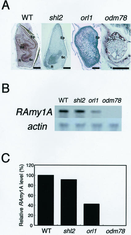
Figure 5.
RAmy1A expression in three types of embryonic organ-deficient mutants. A, Phenotypes of the mature mutant embryos. Each embryo is shown with the embryo-epithelium boundary to the right side. The characteristic phenotype of each mutant is described in the text. SA, Shoot apex; R, root; Sc, scutellum; Ep, epithelium. Bar indicates 500 μm. B, RAmy1A expression in the mutant endosperm. After 96 h of imbibition, total RNA was extracted from the endosperm; 1 μg of total RNA was used for the northern analysis. The lower panel shows a loading control probed with actin cDNA. C, Relative levels of RAmy1A expression in B. The RAmy1A expression levels were normalized against the level of expression of the actin gene. The RAmy1A level in the wild type was set at 100%.

