
Figure 3.
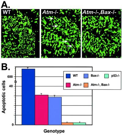
Apoptosis is further reduced in Atm/Bax double-null CNS tissue after IR. (A) Sytox green staining of the P5 cerebellum from WT, Atm-null, and Atm/Bax double-null animals shows a pronounced reduction in the number of apoptotic EGL cells 18 hr after IR in the Atm-null compared with WT. A further reduction was seen in Atm/Bax double-null EGL. Arrows in the Atm−/− section indicate apoptotic cells. (×400.) (B) Quantitative comparison of irradiated cerebellum from various genotypes scored for apoptotic cells. Although the WT cerebellum had abundant cell deaths, the Atm-null and Bax-null cerebellum showed only low numbers of dead cells, whereas the Atm/Bax double-null cerebellum was indistinguishable from the p53-null cerebellum, with an almost complete absence of apoptosis.