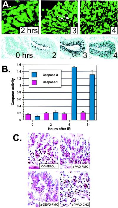
Figure 4.

Caspase-3 activation coincides with apoptosis in the CNS of WT mice. (A) Apoptotic cells appeared between 2 and 3 hr in the cerebellar EGL after IR and by 4 hr, apoptotic figures were widely distributed throughout the EGL. Sections are stained with Sytox green to visualize pyknotic cells; arrows indicate apoptotic cells. (×400.) Immunohistochemistry by using antibodies specific for the cleaved subunit of caspase-3 (15) show caspase-3 activation precedes the apoptotic morphological changes. Caspase-3 cleavage appears initially at 2 hr in the outer EGL, and abundant staining throughout the EGL occurs at 3 and 4 hr. (×200.) (B) Caspase-3 enzyme activity coincides with caspase-3 cleavage and apoptosis seen in A. An increase in caspase-3 activity (pmol of 7-amino-4-methylcoumarin liberated per μg of protein) occurred between 2 and 4 hr after IR in the cerebellum. Caspase-1 activity was insensitive to radiation. (C) Cerebellar slice cultures were irradiated, incubated with various caspase inhibitors, cryosectioned, and stained with neutral red to identify apoptotic cells. Incubation with general caspase inhibitors (z-VAD-FMK) or caspase-3 inhibitor (z-DVED-FMK), but not caspase-1 inhibitor (z-YVAD-CHO) or DMSO vehicle alone (CONTROL), prevented IR-induced apoptosis in the cerebellar EGL. No apoptosis was observed in unirradiated cultures. Sections are from P5 cerebellum. (×400.)
