
Figure 5.
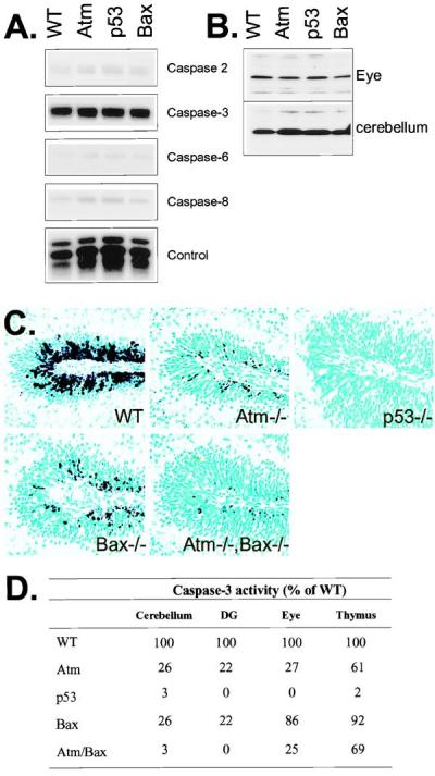
Caspase expression and proteolysis after IR. (A) RPA analysis of the P5 WT, Atm, p53, and Bax-null cerebellum shows a high level of expression for caspase-3 and only low levels for caspases-2, -6, and -8. (B) Protein immunoblot analysis with anticaspase-3 shows equal levels of caspase-3 proenzyme are present in WT, Atm−/−, p53−/−, and Bax−/− tissues. (C) Caspase-3 activation was reduced in the Atm- and Bax-null cerebellar EGL and further reduced in the Atm/Bax double-null animals after irradiation. No caspase-3 activation was observed in the irradiated p53-null cerebellum. (×200.) (D) The relative levels of caspase-3 activity in various mutant tissues 6 hr after IR as a percentage of WT activity. In all cases, samples were normalized for caspase-3 activity present in unirradiated tissues. DG, dentate gyrus.