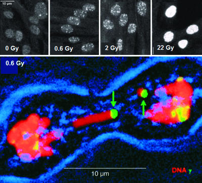
Figure 1.
γ-H2AX foci formation in IMR90 cell cultures and in muntjac mitotic chromosomes. (Upper) IMR90 cell cultures were exposed to the indicated dose from a 137Cs source. After 15 min of recovery at 37°C, the cultures were fixed and processed for immunofluorescence. White dots are γ-H2AX foci. (Lower) Muntjac fibroblast cultures were exposed and permitted to recover for 90 min at 37°C. After processing for immunofluorescence, cultures were scanned through a confocal microscope for mitotic cells. Red, DNA; green, γ-H2AX; blue, phase showing the cell membrane. [Reproduced with permission from ref. 7 (Copyright 1999, The Rockefeller University Press).]

