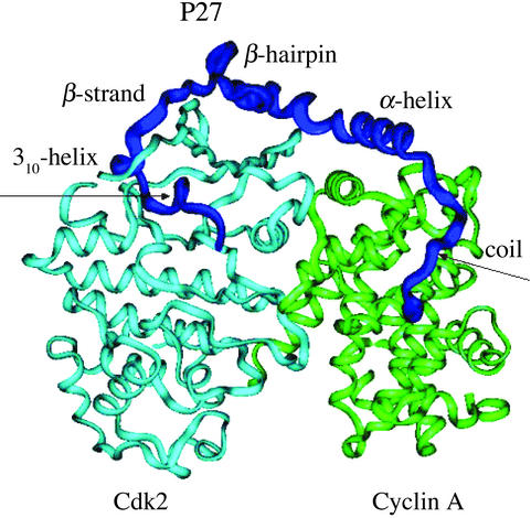
Figure 1.
The crystal structure of the tertiary complex with the bound 69-aa p27 protein (residues 25–93) comprising sequentially the coil (residues from 25 to 34), α-helix (residues from 35 to 60), β-hairpin (residues from 61 to 71), β-strand (residues 75 to 81), and 310-helix (residues from 85 to 90). Schematic drawing shows p27 in blue, cyclin A in green, and Cdk2 in light blue.

