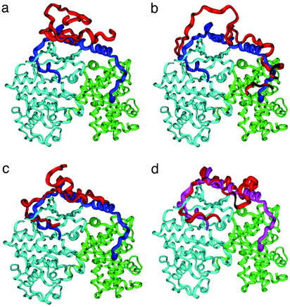
Figure 6.
A schematic representation of the proposed p27 binding mechanism: (a) the ensemble of nonspecific, largely unbound conformations; (b) initiation recruitment of p27 at the cyclin A docking site; (c) the TSE; and (d) the ensemble of postcritical states. The major conformational ensembles (red) are superimposed onto the crystal structure of the bound p27 (blue), cyclin A (green), and Cdk2 (light blue).

