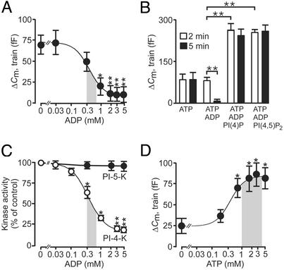
Figure 4.
ADP inhibits exocytosis by suppression of PI 4-kinase activity and RRP replenishment. (A) Exocytosis (ΔCm) elicited by the second of two trains of fourteen 500-ms depolarizations applied 2 and 5 min after establishment of standard whole-cell configuration in the presence 0–5 mM Mg-ADP and 3 mM ATP. The responses to the first train were unaffected by the nucleotide. The curve was drawn by approximating the Hill equation to the mean values. The shaded area highlights the physiological range of ADP concentrations observed at low and high glucose (data taken from 30). (B) Histogram summarizing exocytosis elicited by first (2 min, open bars) and second (5 min after establishing whole-cell configuration, filled bars) train of depolarizations under control conditions (3 mM ATP alone), in the presence of 3 mM ATP and 3 mM ADP, in the presence of 3 mM ATP, 3 mM ADP, and 1 μM of either PI(4)P or PI(4,5)P2. (C) PI 4-kinase (open circles) and PI 5-kinase (filled circles) activities measured in islet homogenates in the presence of 0–5 mM ADP. The curve for PI 4-kinase activity was drawn by approximating the Hill equation to the mean values. The shaded area highlights the physiological range of ADP concentrations observed at low and high glucose (data taken from ref. 30). (D) As in A but the experiment was conducted in the presence of 0–5 mM Mg-ATP. The shaded area indicates the range of ATP concentrations measured in β cells at low and high glucose concentrations (data taken from ref. 30). Data are presented as mean values ± SEM of five experiments. *, P < 0.05; **, P < 0.01.

