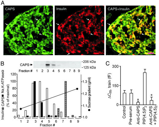
Figure 5.
CAPS is involved in Ca2+- and PI(4,5)P2-dependent exocytosis in β cells. (A) Images of sections of rat endocrine pancreas stained for CAPS (Left) and insulin (Center) and after overlay (Right). CAPS immunoreactivity in single β cells is indicated by arrows. (Scale bar = 20 μm.) (B Upper) Western blot illustrating the presence of CAPS in different subcellular fractions 1–9. (B Lower) Quantitation of the abundance of CAPS (filled bars) and Na+, K+-ATPase α-subunit (plasma membrane fractions, open bars), insulin content (granule-enriched fractions, hatched bars), and the sucrose gradient (black line). The experiment was repeated twice with identical results. (C) Histogram summarizing exocytosis elicited by trains of depolarizations applied 2 min after establishment of the whole-cell configuration under control conditions, in the presence of preimmune serum (preserum; 1.5 mg/ml), after inclusion of an antibody against CAPS (anti-CAPS, 1.5 mg/ml), and the CAPS antibody plus 1 μM of PI(4,5)P2. Data are mean values ± SEM of five experiments. *, P < 0.01.

