Figure 1.
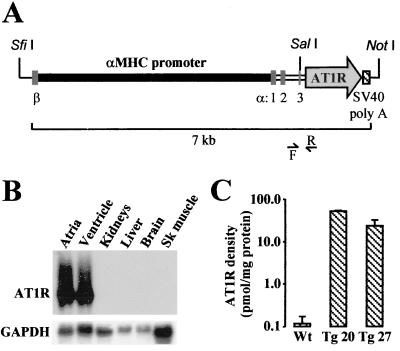
(A) Schematic representation of the αMHC-AT1R transgene. β and α:1, 2, and 3 denote the position of the last exon of βMHC gene and of the three first exons of αMHC gene. F and R indicate the forward and reverse primers used for screening. (B) Cardiac-specific expression of the AT1R transgene. The tissue specificity of AT1R transgene expression was determined in total RNA by Northern blot analysis as described in Methods. Blots were rehybridized with rat glyceraldehyde-3-phosphate dehydrogenase cDNA probe to control for RNA loading. Sk, skeletal. (C) Expression of human AT1R protein. Density of the AT1R was determined in membranes isolated from ventricles of 62- to 140-day-old Wt (n = 6), 62-day-old Tg 20 (n = 3), and 140-day-old Tg 27 (n = 3) mice as described in Methods, using losartan as competitor (data are means ± SEM).

