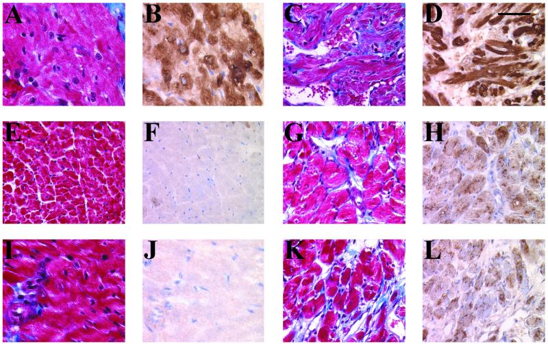
Figure 5.
Increased expression of ANF and collagen deposition in the ventricles of Tg mice. Five-micrometer heart sections from 111-day-old right atria (A and B) and left (E and F) and right (I and J) ventricles of Wt and right atria (C and D) and left (G and H) and right (K and L) ventricles of Tg 27 were immunostained with a rabbit polyclonal anti-ANF antibody and were counterstained with methylgreen (B, D, F, H, J, and L), and collagen deposition (blue) was determined by Masson's trichrome staining (A, C, E, G, I, and K). Note the homogeneously increased ANF immunoreactivity in both Tg ventricles. Interstitial fibrosis is detected in both ventricles and atria of Tg as compared with Wt mice. Significant cardiomyocyte hypertrophy is also evident in Tg ventricles. (Bar = 50 μm.)

